
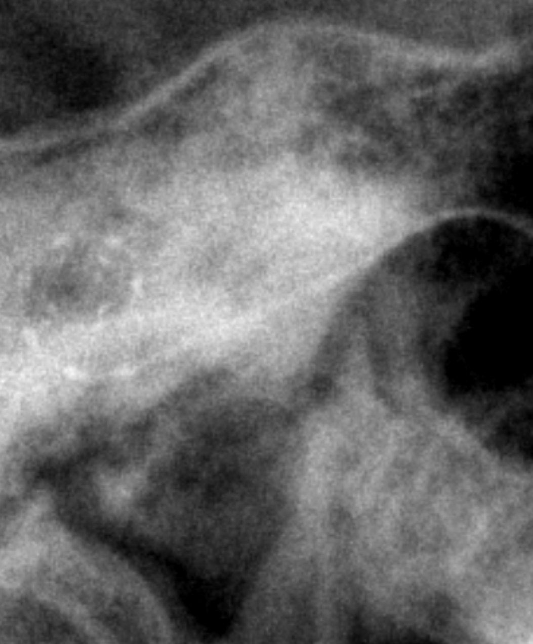
Five days after surgery, I returned to Northwestern for a postoperative CT scan.
This scan wasn’t being done because anyone suspected a problem. It had been planned so the surgical team could see exactly where the electrode array was positioned inside my cochlea. The images would then be sent to an engineer at MED-EL to map the placement of the electrodes. I had been waiting to see this. I got to see the X-Ray of the electrode that was taken during surgery, but I really wanted to see the CT Scan.
After spending weeks researching cochlear implants, and choosing MED-EL largely because my cochlea is longer than average, I wanted to know where that carefully selected electrode had actually landed. The report confirmed that the implant was in place and that the tip of the electrode array terminated at the apical turn of my cochlea. In other words, the electrode had traveled deeply through the spiral and reached the innermost portion.
This was exactly the kind of information the MED-EL engineer needed. By identifying where the individual electrode contacts were positioned within my cochlea, the team could better understand which areas each contact would stimulate when my implant was activated and mapped. The CT also showed plenty of evidence that someone had recently drilled into my skull. There was fluid, gas and tissue debris around the surgical area, along with opacification in the left mastoid and middle ear. It sounds alarming when written in radiology language, but these were described as evolving postoperative changes.
They also helped explain why my ear still felt so full and pressurized. The important surrounding structures including the ossicles, bony labyrinth, internal auditory canal and facial nerve canal remained intact. I love when they send me this stuff.
There is something fascinating about reading the report and seeing physical evidence of what was done inside my head. Until then, the implant was mostly something I understood conceptually. I knew it was there, of course, but I couldn’t see it or use it yet. Now I could see the electrode curling through my cochlea. Physically, however, I was still very much in the postoperative-healing stage. My ear had settled into a repeating pattern: Gentle throbbing. Stabby pain. Rinse and repeat.
Nothing appeared infected or alarming, but my ear was making it very clear that surgery had occurred only five days earlier. Naturally, that was also the day my new bed desk arrived. Joe set it on the bed so I could assemble it, because apparently neither cochlear implant surgery nor intermittent stabby head pain could suppress my need to put things together.
The CT gave us the information we needed. The electrode was in place. The images were headed to MED-EL for analysis. Now there was only one major thing left to do: Turn it on.

Comments